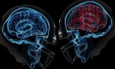
Finding Football-Related Brain Damage with MRI
Florida researchers report that over 40% of the retired NFL football players they studied had signs of brain damage1. This sad and shocking statistic was the result of using diffusion-tensor MRI sequences, a specific “tuning” of magnetic resonance imaging that enhances diagnosis of brain problems.
Typical MRI brain scans on a powerful 3-Tesla (3T) magnet use diffusion weight imaging (DWI) that shows abnormal areas by depicting the restriction of water molecule motion in injured tissue. It provides qualitative and quantitative visual information on tissues and fluids that indicate swelling, unusual bleeding, etc. DWI has been used with MR scans since the mid-1980s2. More recently, thanks to advances in software and hardware, a new functional sequence called diffusion-tensor imaging (DTI) makes it possible to analyze microstructures in the brain with high sensitivity. Using DWI and DTI together gives crucial information at the earliest possible time to maximize the effectiveness of interventions. Put another way, you don’t want a brain injury to hang around and get worse.
A research team at the Florida Center for Headache and Sports Neurology scanned 40 former NFL players over a period of several years in order to “evaluate them for memory loss, chronic headaches, and other neurological abnormalities3.” One of the study authors, Dr. Francis Conidi, was astonished at the high numbers. The implications are that the longer an athlete is involved in his sport, the greater the risk for brain injury.
Along with DTI, other imaging sequences were used. The combination of all scan results showed evidence of nerve damage as well as injury to the brain itself. The players were also given cognitive and psychological tests, another way to look for diminished mental and emotional ability. In many cases, these tests also showed the lingering aftereffects of the kinds of head trauma that football players can suffer.
The study underscores the importance of identifying brain trauma as soon as possible after a head injury. The Sperling Diagnostic Center provides state-of-the-art head and spinal cord imaging, with a sophisticated magnet that can be readily adapted to scan even very young children who experience an accident or illness that affects the brain and nervous system. When problems are identified early, treatments for head injury have the highest success. In this way and in many others, our Diagnostic Center is a source of hope and healing.
_______________________________
1 Forrest, Wayne. “MRI shows brain injury in nearly half of ex-NFL players.” April 11, 2016. http://www.auntminnie.com/index.aspx?sec=sup&sub=mri&pag=dis&itemid=113922
2 Huisman, TA. Diffusion-weighted and diffusion tensor imaging of the brain, made easy. Cancer Imaging. 2010; 10(1A): S163–S171. http://www.ncbi.nlm.nih.gov/pmc/articles/PMC2967146/
3Forrest, ibid.
- CATEGORY:
- Neurological MRI